BHB resumes biopsies with its new 3D mammography machine

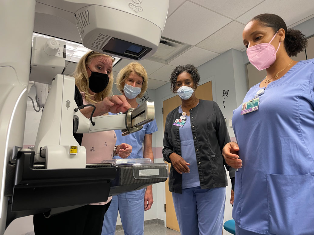
Wednesday 9 June 2021: Bermuda Hospitals Board (BHB) is pleased to announce the resumption of stereotactic biopsy (biopsies that are carried out using our mammography machine) service today. The recent upgrade to the GE Pristina 3D mammography unit meant these types of biopsies had to be delayed until staff were fully trained on the new machine. That training has started, and today the first patients received their biopsies using the new equipment.

Imaging Services staff are excited about the upgrade and training, and the improvement this brings to their patients.

The first patient today, a 54-year-old woman, said she was pleased to have the procedure on state-of-the-art equipment.

She said:  “It was great. The staff were wonderful, they fully explained the procedure and I didn’t feel a thing. I was comfortable and relaxed and now am eager to get the results. I’m staying positive that it will be good news.”

“In diagnostic imaging, image clarity is what we get excited about,” said Chief of Radiology Daniel Stovell, MD. “The clearer the image, the better our ability to detect, diagnose and treat any abnormalities.”

BHB is the only provider of 3D stereotactic breast biopsy in Bermuda at this time.

9 June 2021 Home Page, News

BHB will introduce automated breast ultrasound service

Thursday 22 April 2021: Bermuda Hospitals Board is upgrading its mammography service with a new 3D unit and possibly the island’s first automated breast ultrasound unit (ABUS).

The Pristina 3D mammography unit and the Ivenia ABUS, both from manufacturer GE, will be installed this month. To make room for the new units, the old 2D mammography machine is being removed.

Mammography and biopsy services have been suspended while the switchover is carried out. Following the installations, staff training on the new equipment will take place.

Benefits of 3D mammography

Research has shown that 3D mammography locates more cancers than its 2D predecessor and also reduces the number of false positives. In 2D mammography, two X-ray images of the breast are used. Three-dimensional (3D) mammography creates a 3D picture of breast tissue using X-ray images of several different angles around the breast.

Benefits of automated breast ultrasound

In mammograms, both the healthy dense breast tissue and cancer appear white. This makes cancer detection more difficult in those with dense breasts. Seventy-one percent of cancers occur in dense breasts, and studies show that over a third of cancers in dense breasts are missed in mammograms.

Cancer detection in dense breast tissue significantly improves with the use of a specially designed ultrasound device – ABUS. The combination imagery of X-rays (from the 3D mammogram) and sound waves (from the ultrasound) produces a much clearer picture of the dense breast tissue. Cancer cells appear black in ultrasound imagery, while the dense breast tissue appears white. This makes it easier for radiologists to detect cancer cells.

“Imaging Services staff are excited about this upgrade and eager to start using the advanced technology,” said Diagnostic Imaging Clinical Manager Renee Butterfield.

“The installations represent significant improvement in service we provide the public,” said Chief of Diagnostic Imaging Daniel Stovell, MD. “The technologies produce clearer images, which have a host of medical benefits, one of the most important of which is that it improves our ability to detect cancer.”

22 April 2021 Home Page, News